ERG test: Advancements in eye exams to test for plaquenil toxitity right around the corner!
As far as tests go, the ERG is not one of my favorites. The ERG, or Electroretinography, is one of those tests that seems to last way too long and comes up way too fast. When the scheduled appointment comes up on my calendar, I'm find myself thinking, "Didn't I just have one of these?"
Of course, like many of the tests that we have for lupus, and the side-effects of the medication we take for it, (of which the ERG is one), it's a necessary evil. The test is done to detect any changes in the retina, in my case, due to the use of Plaquenil. My understanding is that it's not months on this drug that can cause changes, but rather, years.
I was on plaquenil for three years the first time around, before my opthamalogist detected some changes and recommended that I stop using the drug. It's hard to know what exactly those changes were, and if they were indeed from plaquenil, but my rheumatologist and I didn't question it. The opthamalogist saw something he didn't like, so we stopped using the medication. A few years later, I started the medication again, and have been successfully taking it for the past seven years, with no retinal changes detected at all.
But, like the rest of you who take Plaquenil, I do what I need to do to take care of my eyes. I visit my opthamalogist every six months to a year for regular checkups, and to take a visual field test (which happens to be another non-favorite of mine). And for the past few years, I've been making annual visits to a specialized opthamalogist who administers the ERG.
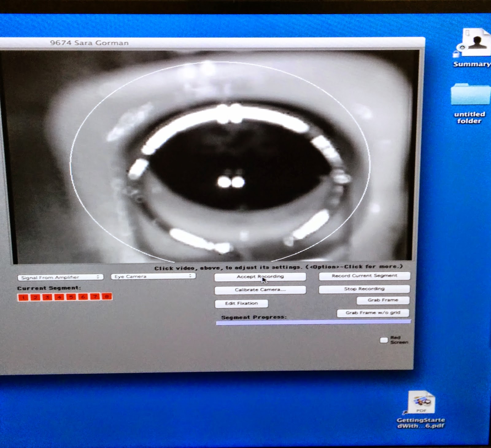
The last time I was in, I asked the nurse to take a picture of the computer screen that was recording the visual of my eye during the test. I hope this doesn't freak you out. If you remember, I can hardly even look at pictures of my own rashes, but this doesn't bother me. Probably because it doesn't even look like my eye - but rather, a ship from outerspace!
When my eye looked like this, my eyes were propped open with something like a thick contact lens, although it felt more like a big old monocle to me. Here's exactly what was happening:
 The best news to come out of my test this year? They're very close to administering this test a different way!
Below you can read a little bit more about an ERG. Enjoy!
Of course, like many of the tests that we have for lupus, and the side-effects of the medication we take for it, (of which the ERG is one), it's a necessary evil. The test is done to detect any changes in the retina, in my case, due to the use of Plaquenil. My understanding is that it's not months on this drug that can cause changes, but rather, years.
I was on plaquenil for three years the first time around, before my opthamalogist detected some changes and recommended that I stop using the drug. It's hard to know what exactly those changes were, and if they were indeed from plaquenil, but my rheumatologist and I didn't question it. The opthamalogist saw something he didn't like, so we stopped using the medication. A few years later, I started the medication again, and have been successfully taking it for the past seven years, with no retinal changes detected at all.
But, like the rest of you who take Plaquenil, I do what I need to do to take care of my eyes. I visit my opthamalogist every six months to a year for regular checkups, and to take a visual field test (which happens to be another non-favorite of mine). And for the past few years, I've been making annual visits to a specialized opthamalogist who administers the ERG.
The last time I was in, I asked the nurse to take a picture of the computer screen that was recording the visual of my eye during the test. I hope this doesn't freak you out. If you remember, I can hardly even look at pictures of my own rashes, but this doesn't bother me. Probably because it doesn't even look like my eye - but rather, a ship from outerspace!
When my eye looked like this, my eyes were propped open with something like a thick contact lens, although it felt more like a big old monocle to me. Here's exactly what was happening:
How an ERG is done:
The patient assumes a comfortable position (lying down or sitting up). Usually the patient's eyes are dilated beforehand with standard dilating eye drops. Anesthetic drops are then placed in the eyes, causing them to become numb. The eyelids are then propped open with a speculum, and an electrode is gently placed on each eye with a device very similar to a contact lens. An additional electrode is placed on the skin to provide a ground for the very faint electrical signals produced by the retina.
During an ERG recording session, the patient watches a standardized light stimulus, and the resulting signal is interpreted in terms of its amplitude (voltage) and time course. The visual stimuli include flashes, called a flash ERG, and reversing checkerboard patterns, known as a pattern ERG.
The electrodes measure the electrical activity of the retina in response to light. The information that comes from each electrode is transmitted to a monitor where it is displayed as two types of waves, labeled the A waves and B waves.
After we did this test with the monocle, I mean, contact lens, in my eye, the nurse administered the entire test a second time, this time merely taping a thin film over my eye. No propping, no monocle, no discomfort! My doctor is hopeful that by the next time I have the test done, they will have had enough data compiled (hence the reason I did the test twice) to determine that the thin film is just as effective as that big old contact lens. My fingers are seriously crossed. But it was exciting to be on the cusp of such technological advances in medicine!
Along those same lines, a frequent reader of my blog was kind enough to share another advancement in eye procedures - this time in regard to cataract surgery. You can read more about that here. Thanks, Jen, for sharing!Below you can read a little bit more about an ERG. Enjoy!
What is electroretinography?
Electroretinography (ERG) is an eye test used to detect abnormal function of the retina (the light-detecting portion of the eye). Specifically, in this test, the light-sensitive cells of the eye, the rods and cones, and their connectingganglion cells in the retina are examined. During the test, an electrode is placed on the cornea (at the front of the eye) to measure the electrical responses to light of the cells that sense light in the retina at the back of the eye.
Why is an ERG done?
An ERG is useful in evaluating both inherited (hereditary) and acquired disorders of the retina (like plaquenil retinal toxicity). An ERG can also be useful in determining if retinal surgery or other types of ocular surgery such as cataract extraction might be useful.


Comments
If you are looking for Online Doctor Appointment, NamasteDoc is a portal that enables online consultation with doctors in India. It connects patients to doctors online and manages appointments effortlessly. Book appointments anytime, anywhere as per your convenience.
http://www.namastedoc.com/
http://www.optometricretinasociety.org/
and the paper on New Plaquinil Guidelines.